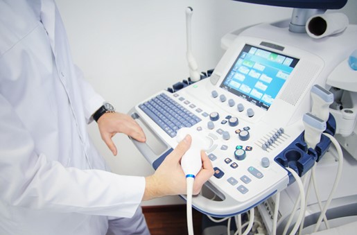
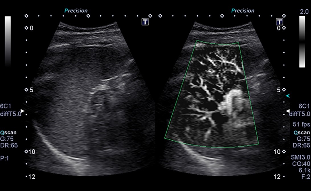
Ultrasound imaging is a term that almost everyone has heard at one point or another. There are many reasons why diagnostic ultrasound is one of the most common testing processes done in the medical field. Ultrasound scanning is so common that you will find a diagnostic ultrasound machine in every medical testing facility. However, while people know what an ultrasound examination is, not many have an in-depth understanding of how it works.

What is Medical Ultrasound Imaging?
It is a medical imaging method that uses sound waves on a body’s internal organs for testing, diagnostic, or therapeutic reasons. The sound waves travel through the body and are converted into an ultrasound image showing the condition and boundaries of fluid and soft tissue and internal organs in the body. This allows medical staff to diagnose problems and decide on treatment programs. Using medical ultrasound imaging allows doctors to diagnose problems with internal organs and sources of inflammation or pain in the body. Not only that, but ultrasound imaging is the most common testing method used on pregnant women to monitor the growth of a fetus inside the body.This imaging technique uses ultrasound waves, which are very high-frequency sound waves. These sound waves cannot be heard or differentiated by human ears.

What is the difference between Ultrasound and Sonography?
Ultrasound is a noninvasive, painless procedure that uses high-frequency sound waves to produce images of organs, soft tissues, blood vessels, and blood flow, from inside the body. These images are used for medical analysis. After x-ray exams, ultrasound is the most commonly used form of diagnostic imaging. It helps doctor gain insights into the inner working of the body, and is known for being safe, radiation-free, noninvasive, portable, widely accessible, and affordable. Sonography, on the other hand, is the use of an ultrasound tool for diagnostic purposes. Medical sonographers, often referred to as ultrasound techs, are the people trained to use ultrasound diagnostic imaging technology (sonography). They provide doctors with detailed images of what’s going on inside of patients.
What is the difference between Ultrasound and CT Scan?
A Computerized Tomography (CT) scan is another common medical imaging procedure used to identify ailments inside the body. However, this is quite different from ultrasound scan procedures.
CT scans utilize X-rays to generate a detailed image of the body’s internal organs. The X-ray tube rotates to capture images of different body sections and tissues.
Since CT scans use X-rays, the procedure is radioactive and potentially harmful to some extent. However, ultrasound scans produce no ionizing radiation exposure since they use sound waves instead.


